Introduction
Breast cancer remains one of the most common malignancies affecting women worldwide, with an estimated 2.3 million new cases diagnosed in 2020.1 In recent years, neoadjuvant chemotherapy (NAC) has emerged as a standard treatment…
Breast cancer remains one of the most common malignancies affecting women worldwide, with an estimated 2.3 million new cases diagnosed in 2020.1 In recent years, neoadjuvant chemotherapy (NAC) has emerged as a standard treatment…

The People’s Republic of China’s Zhang Qingying and Japan’s Sugihara Aiko won the women’s balance beam and floor exercise titles, respectively, on Saturday (25 October) on the final day of competition at the 2025 World Gymnastics Championships in…
Asthma is a heterogeneous respiratory disease, generally classified into Type 2 (T2) and non-Type 2 (non-T2) subtypes, characterized by chest tightness, cough, wheezing, and shortness of breath, affecting approximately 1–18% of the global population.1–3 Previous studies have shown that airway epithelial damage plays a crucial role in asthma pathogenesis.4,5 Despite continual advancements in clinical guidelines and therapeutic strategies, a substantial proportion of patients fail to achieve adequate disease control. This leads to frequent exacerbations and progressive decline in pulmonary function,6 underscoring the urgent need for highly sensitive and specific biomarkers to enhance diagnostic accuracy and optimize management strategies. Ferroptosis is an iron-dependent form of regulated cell death driven by lipid peroxidation,7 which can trigger inflammatory cascades through the release of damage-associated molecular patterns (DAMPs) like reactive oxygen species (ROS).8 Earlier studies have shown that ferroptosis in airway epithelial cells contributes to the inflammatory response and plays a key role in the pathological progression of asthma.9,10 Nevertheless, it remains unclear whether ferroptosis-related genes (FRGs) have diagnostic or therapeutic value in asthma.
The CRYAB gene encodes αB-crystallin, a small heat shock protein that functions as a molecular chaperone and modulates multiple intracellular signaling pathways, thereby influencing cellular proliferation and apoptosis.11,12 Emerging evidence suggests that novel therapeutic strategies targeting CRYAB or its interacting proteins—by selectively inducing apoptosis or disrupting metastatic processes—can improve outcomes in patients with metastatic disease.13,14 Although CRYAB has been implicated in the pathogenesis and progression of various disorders, its specific role in asthma remains unclear, as does its involvement in the regulation of ferroptosis.
Machine learning algorithms such as LASSO and SVM have shown significant potential in identifying disease biomarkers by selecting key feature genes from large datasets.15,16 Zhang et al found that STUA1 and SLC27A3 are valuable diagnostic biomarkers for chronic obstructive pulmonary disease (COPD) and affect the mechanism of COPD to a certain extent through the combined LASSO and SVM algorithms.17 Previous studies have demonstrated that exposure to house dust mites disrupts systemic iron homeostasis, exacerbates lipid peroxidation, and activates ferritinophagy in asthma, ultimately inducing ferroptosis in airway epithelial cells and amplifying inflammatory responses.18 Consistent with these findings, both asthma animal models and cell models exhibit markedly elevated levels of Fe²⁺, malondialdehyde (MDA), and reactive oxygen species (ROS), accompanied by reduced glutathione (GSH) levels—hallmarks of enhanced ferroptosis—which further accelerate asthma progression.19 Based on this body of evidence, we hypothesize that CRYAB, as a pivotal component of the ferroptosis regulatory network, may play a critical role in the pathophysiological processes of asthma. In the present study, we employed an integrated approach combining LASSO and SVM algorithms to systematically identify FRGs associated with asthma from the GEO database. Candidate FRGs were subsequently validated using clinical specimens, followed by functional experiments involving CRYAB overexpression or silencing in airway epithelial cells to elucidate its biological roles. Collectively, our findings not only reveal a potential mechanistic link between FRGs-particularly CRYAB-and immune regulatory networks but also provide a theoretical framework for developing precision diagnostic biomarkers as well as novel therapeutics targeting ferroptosis-related pathways for improved management of asthma.
The GEO (Gene Expression Omnibus) database (https://www.ncbi.nlm.nih.gov/geo/) is an open-access repository that archives high-throughput gene expression data submitted by research groups worldwide.20 Based on our research objectives, we identified and included three microarray datasets for further analysis: GSE137268 (54 asthma patients and 15 healthy controls), GSE148004 (9 asthma patients and 10 healthy controls), and GSE4302 (42 asthma patients and 28 healthy controls). We grouped these datasets as follows: GSE137268 and GSE148004 were designated as the experimental cohorts for mining disease-specific gene expression profiles, while GSE4302 served as an independent validation cohort to assess the accuracy of our findings.
In this study, we used the “Limma” package in R to identify DEGs. The screening criteria were set as follows: |Log2 Fold Change (Log2FC)|> 0.5, and an adjusted P-value (adjust P.Val) < 0.05. Initially, we compared asthma samples with healthy controls within the experimental group to obtain the set of DEGs.
Disease Ontology (DO) enrichment analysis was conducted using the DOSE package to further investigate disease mechanisms and support novel drug development,21 with p < 0.05 set as the threshold for significance.
FRGs were downloaded from the FerrDb database and intersected with the previously identified DEGs to generate a list of candidate diagnostic biomarkers. Feature selection was performed using LASSO and SVM-RFE algorithms to identify potential diagnostic biomarkers from asthma patients, with p < 0.05 considered statistically significant. The predictive value of these FRGs for asthma was assessed using receiver operating characteristic (ROC) curve analysis, and their diagnostic performance was further validated using the GSE4302 dataset.
To investigate the role of immune cells in the pathogenesis of asthma, we performed single-sample Gene Set Enrichment Analysis (ssGSEA) using the “GSVA” package in R.
For this study, lung lobe and segmental bronchial specimens were collected from patients undergoing lobectomy who met the inclusion criteria. A total of 12 specimens were obtained, including 6 from non-asthmatic patients and 6 from asthmatic patients. All participants provided written informed consent prior to sample collection. The study protocol was approved by the Ethics Committee of Henan Provincial People’s Hospital (No. 2024–077-01).
The human bronchial epithelial cell line BEAS-2B was purchased from FuHeng (FH0319, Shanghai, China). Cells were cultured in DMEM supplemented with 10% fetal bovine serum and 1% penicillin-streptomycin in a 37°C, CO2 incubator.
Total protein was extracted from lung tissue or cells using RIPA lysis buffer (Jingcai Bio, Xian, China) and separated by sodium dodecyl sulfate-polyacrylamide gel electrophoresis (SDS-PAGE). Subsequently, protein bands in the gel were transferred onto polyvinylidene difluoride (PVDF) membranes. The membranes were blocked with 5% non-fat milk and incubated with primary anti-CRYAB overnight at 4°C, followed by incubation with IRDye 680RD Goat anti-Rabbit for 1 hour at room temperature. The relative expression of protein was measured using Image J, with β-actin/GAPDH serving as the loading control.
Total RNA was isolated from human lung tissue or cells using TRIzol reagent (Invitrogen, USA) according to the manufacturer’s protocol. Use a reverse transcription kit (Servicebio, Wuhan, China) to reverse transcribe RNA into cDNA. RT-qPCR was carried out on an ABI 7500 qPCR system using the 2× SYBR Green qPCR Master Mix (Vazyme, Nanjing, China) reagent. The endogenous reference gene primers for β-actin, as well as other RT-qPCR primers, are listed in Table 1, all of which were obtained from Sangon Biotech (Shanghai, China). All experiments were repeated three times. The relative expression level was calculated using the 2−ΔΔCt method, with β-actin mRNA serving as an internal control for normalization.
|
Table 1 Primers Used in the RT-qPCR Reaction
|
When cells reached 50–70% confluence in complete growth medium, they were transfected with either siRNAs or plasmids. The siRNAs and pcDNA3.1 constructs were synthesized by Sangon Biotech (Shanghai, China). Ferroptosis was induced by exposing the transfected cells with 10 μM erastin (MCE, USA) for 24 hours. All experiments were performed 48 hours after transfection.
Intracellular ROS levels were measured using the ROS Assay Kit (Solarbio, Beijing, China). BEAS-2B cells were seeded at a density of 2×10⁵ cells per well in a 12-well plate. Following transfection with siRNA or pcDNA3.1 for 24 hours, cells were washed and incubated with PBS. The medium was then replaced with 500 μL of working solution containing 10 μM H2DCFDA, and the cells were incubated at 37°C for 30 minutes. After washing twice with serum-free medium, ROS fluorescence was visualized and imaged using a laser confocal microscope (ZEISS LSM800, Oberkochen, Germany).
BEAS-2B cells were seeded at a density of 5×10³ cells per well in a 96-well plate. After transfection with siRNA or pcDNA3.1 plasmids, cell viability was assessed at 24, 48, and 72 hours using the CCK-8 assay (MCE, USA). CCK-8 solution was added to each well and incubated for an additional 1.5 hours. Absorbance was then measured at 450 nm using a microplate reader (Thermo Scientific, USA).
Reduced GSH levels were measured using the GSH Content Assay Kit (Jiancheng, Nanjing, China), and MDA levels were assessed with the Lipid Peroxidation MDA Assay Kit (Beyotime, Shanghai, China), following the manufacturers’ protocols. Cell preparation was performed as described for ROS detection.
All data are presented as mean ± standard deviation (SD) and were analyzed using GraphPad Prism 9.0 software (USA). Comparisons between two groups were made using an unpaired Student’s t-test; for variables not conforming to a normal distribution, the Mann–Whitney test was applied. p < 0.05 was considered statistically significant.
After performing differential analysis on the experimental group dataset, a total of 213 DEGs were identified, including 39 downregulated and 174 upregulated genes. These DEGs were further visualized using heatmaps and volcano plots, as shown in Figure 1a and b. DO analysis refers to the enrichment analysis of differentially expressed or target gene sets using the DO database, in order to elucidate their potential roles in diseases such as asthma and other respiratory disorders.22 The results of this analysis (Figure 1c and d) indicated that the primary diseases significantly enriched among these DEGs included asthma, obstructive pulmonary disease, bronchial disease, and other respiratory system diseases. Therefore, this study preliminarily suggests that these DEGs are closely associated with the occurrence and development of asthma, thereby providing new insights into its diagnosis and treatment.
 |
Figure 1 Analysis of DEGs and disease ontology enrichment in asthma samples. (a) Heatmap of DEGs. (b) Volcano map of DEGs. (c) Bar chart of DO analysis results. (d) Bubble chart of DO analysis results.
|
To investigate the role of ferroptosis in the pathogenesis of asthma, we retrieved a list of FRGs from the FerrDb database. Differentially expressed FRGs were identified using Venn diagram analysis, resulting in five candidate genes: TFRC, ACO1, CRYAB, SLC7A11, and GCLM (Figure 2a). To further determine which of these may serve as asthma-specific signature genes, we employed two machine learning algorithms: LASSO and SVM-RFE. LASSO regression identified four hub genes (TFRC, CRYAB, SLC7A11, and GCLM), while SVM-RFE selected five genes (TFRC, ACO1, CRYAB, SLC7A11, and GCLM), To enhance specificity, the intersection of both algorithms was taken. Ultimately, four FRGs—TFRC, CRYAB, SLC7A11, and GCLM—were identified as potential signature genes for asthma, as shown in Figure 2b–d.
 |
Figure 2 Analysis results of FRGs in asthma. (a) Venn diagram showing the identification of differentially expressed FRGs. (b) LASSO regression analysis plot. (c) SVM-RFE algorithm analysis plot. (d) Venn diagram depicting the final selection of signature genes.
|
To determine the specificity and diagnostic value of the identified signature FRGs for asthma, ROC curve analysis was performed to assess their performance. The results showed (Figure 3a–d) that the areas under the curves (AUCs) of these four signature genes were 0.767 (CRYAB), 0.726 (GCLM), 0.722 (SLC7A11), and 0.756 (TFRC). Notably, both CRYAB and TFRC exhibited AUC values greater than 0.75, suggesting superior clinical utility as diagnostic biomarkers in asthma. Furthermore, we analyzed the expression levels of TFRC and CRYAB in an independent validation cohort (GSE4302). The results revealed CRYAB expression was significantly upregulated in patients with asthma, whereas TFRC expression was significantly downregulated compared to controls (Figure 3e and f). To further validate their diagnostic potential, ROC analyses were conducted based on their expression levels in the validation group, and AUC values were calculated as 0.659 for CRYAB and 0.629 for TFRC (Figure 3g and h). Taken together, results from both discovery and validation cohorts indicate that altered expression levels of CRYAB and TFRC may serve as promising diagnostic biomarkers or potential therapeutic targets for asthma.
 |
Figure 3 Diagnostic value and validation of signature FRGs in asthma. (a–d). ROC curve graphs of the diagnostic efficacy of CRYAB, GCLM, SLC7A11, and TFRC for asthma. (e and f). Bar plots showing the expression levels of CRYAB and TFRC. (g and h). ROC curve graphs of the diagnostic efficacy of CRYAB and TFRC for asthma in the validation dataset. Notes: AUC > 0.7 is considered indicative of acceptable diagnostic value. Con = healthy control samples, Treat = asthma samples.
|
Immune cells play a crucial role in the pathological process of asthma, particularly in mediating inflammatory responses and airway remodeling. To investigate differences in immune cell infiltration between asthmatic patients and healthy controls, ssGSEA was performed on the experimental dataset. As shown in Figure 4a, the analysis revealed significantly increased infiltration of activated B cells, activated dendritic cells, myeloid-derived suppressor cells (MDSCs), macrophages, and mast cells in the peripheral blood of asthma patients compared to controls.
 |
Figure 4 Analysis of immune cell infiltration. (a) Diagram of the differences in the infiltration of 28 types of immune cells between asthma and normal control samples. (b) Correlation diagram of CRYAB and TFRC with the abundance of immune cell infiltration. *p<0.05, **p<0.01, ***p<0.001.
|
To further elucidate their roles, we analyzed correlations between these hub genes and immune cell abundance. The results demonstrated that CRYAB expression was positively correlated with plasmacytoid dendritic cells, neutrophils, mast cells, eosinophils, activated dendritic cells, activated CD4 T cells, and activated B cells. Additionally, TFRC expression showed a positive correlation with immature dendritic cells but negative correlations with monocytes, macrophages, effector memory CD8 T cells and activated B cells (Figure 4b). Collectively, these findings suggest that CRYAB and TFRC may influence asthma pathophysiology through modulation of immune cell activity.
To further verify the differential expression of CRYAB in clinical samples, we assessed its mRNA and protein levels in lung tissues from asthma patients and non-asthmatic controls using RT-qPCR and WB, respectively. As shown in Figure 5a and b, CRYAB expression was significantly upregulated in the asthma group compared to controls, while TFRC expression showed no significant difference between groups. Consistently, Western blot analysis confirmed increased CRYAB protein levels in asthmatic lung tissues (Figure 5c and d). As CRYAB is a FRG implicated in asthma, we speculate that it may contribute to disease pathogenesis by modulating ferroptosis.
 |
Figure 5 Expression of CRYAB in patients with asthma and non-asthma controls. (a) Histogram of CRYAB mRNA expression in the two groups. (b) Histogram of TFRC mRNA expression in the two groups. (c and d) Representative WB analysis and quantification of CRYAB protein expression in both groups. **p<0.01, ***p<0.001.
|
CRYAB, a key heat shock protein, is known to confer protection against oxidative stress-induced apoptosis, but its impact on ferroptosis in BEAS-2B cells remains unknown. To elucidate this, we performed siRNA-mediated knockdown and pcDNA3.1-mediated overexpression of CRYAB in BEAS-2B cells. RT-qPCR and Western blot analyses demonstrated effective knockdown by all three siRNAs (si-CRYAB-1/2/3), with si-CRYAB-3 exhibiting the highest silencing efficiency and thus selected for subsequent experiments (Figure 6a–c). Conversely, transfection with pcDNA3.1-CRYAB significantly increased both mRNA and protein levels of CRYAB compared to negative control (Figure 6d–f). Furthermore, erastin-induced ferroptosis resulted in a significant downregulation of CRYAB expression at both mRNA and protein levels (Figure 6g–i).
 |
Figure 6 Expression of CRYAB during ferroptosis. (a–c) siRNA-mediated knockdown of CRYAB significantly reduced its mRNA and protein expression levels. (d–f) pcDNA3.1-mediated overexpression markedly increased CRYAB mRNA and protein expression. (g–i) Downregulation of CRYAB mRNA and protein expression during ferroptosis induction. **p<0.01, ***p<0.001.
|
To investigate whether CRYAB participates in ferroptosis by affecting the lipid peroxidation and antioxidant systems in human bronchial epithelial cells, we employed reagent kits to measure the levels of MDA and GSH. As shown in Figure 7a–d, erastin significantly elevated MDA levels while reducing GSH content in BEAS-2B cells, similar changes were observed upon CRYAB knockdown. In contrast, overexpression of CRYAB decreased MDA accumulation and increased GSH levels relative to controls. Additionally, intracellular ROS levels and cell viability were evaluated: overexpression of CRYAB reduced ROS accumulation and improved cell viability compared to the silenced group, which exhibited enhanced ROS production and decreased viability (Figure 7e–h). Collectively, these results suggested that CRYAB upregulation attenuates ferroptosis by maintaining redox homeostasis and reducing lipid peroxidation in airway epithelial cells. This highlights its potential protective role in the pathogenesis of asthma through regulation of ferroptotic pathways.
 |
Figure 7 Effects of CRYAB knockdown or overexpression on ferroptosis-related markers in BEAS-2B cells. (a and b) Bar charts of GSH and MDA levels following CRYAB knockdown. (c and d) Bar charts of GSH and MDA levels following CRYAB overexpression. (e) Immunofluorescence images showing ROS accumulation after modulation of CRYAB expression. (f and g) Histograms of ROS immunofluorescence intensity. (h) Line charts of relative viability of BEAS-2B cells with altered CRYAB expression. *p<0.05, **p<0.01, ***p<0.001.
|
Asthma is one of the most common chronic diseases worldwide, with a continuously increasing prevalence and affecting over 300 million people globally. Regardless of economic development status, asthma remains a significant public health challenge in most countries.23–25 The pronounced heterogeneity of asthma often limits the effectiveness of conventional treatments; therefore, guiding diagnostic and therapeutic decisions based on specific molecular mechanisms, rather than relying solely on clinical manifestations, has become a central tenet of contemporary precision medicine. Ferroptosis, an iron-dependent form of regulated cell death, has recently been demonstrated to be closely associated with asthma.26,27 Additionally, algorithmic tools such as LASSO and SVM-RFE have provided greater convenience for disease analysis.28 In this study, we sought to explore the diagnostic value of FRGs in asthma and determine their expression levels in asthma patients as well as their role in ferroptosis within bronchial epithelial cells, thereby providing new biomarkers and drug targets for the diagnosis and treatment of asthma.
In this study, we identified 213 DEGs in the asthma group, with upregulated genes predominating. DO enrichment analysis further demonstrated that these DEGs are significantly associated with respiratory diseases, particularly asthma, obstructive pulmonary disease, and bronchial disorders. These findings underscore their potential involvement in the pathogenesis of asthma. To further elucidate specific mechanisms underlying asthma development, we focused on FRGs, given growing evidence linking ferroptosis to asthma. Using LASSO and SVM-RFE algorithms, we narrowed down potential diagnostic biomarkers to four hub FRGs: TFRC, CRYAB, SLC7A11, and GCLM. Previous studies have demonstrated that TFRC, CRYAB, SLC7A11, and GCLM play pivotal regulatory roles in key biological processes of ferroptosis, particularly in the regulation of intracellular iron metabolism and maintenance of redox homeostasis.12,29–31 The identification of these FRGs as putative signature genes for asthma not only provides novel insights into disease mechanisms but also offers promising targets for biomarker development or therapeutic intervention. This lays the foundation for future clinical validation and functional studies aimed at improving diagnosis and management strategies for patients with asthma.
Our study systematically evaluated the diagnostic potential of four FRGs in asthma. Among them, TFRC and CRYAB exhibited consistent expression patterns and demonstrated significant diagnostic value. Specifically, CRYAB was significantly upregulated while TFRC was downregulated in asthma patients compared to controls. These results suggest that altered expression of CRYAB and TFRC may serve as reliable molecular biomarkers for asthma diagnosis across diverse patient populations and implicate their involvement in ferroptosis-associated disease mechanisms. Previous research has shown that activation of TFRC mediates ROS generation and promotes cellular iron accumulation; conversely, inhibition of the TFRC pathway can suppress lung cancer cell growth, indicating its potential as a therapeutic target.32,33 Additionally, high CRYAB protein expression is clinically and pathologically relevant to the disease, including TNM stage and overall survival rate, playing a crucial role in the biology of non-small cell lung cancer.14 In summary, our findings highlight CRYAB and TFRC as promising candidate biomarkers or therapeutic targets related to ferroptosis pathways underlying asthma pathogenesis. Notably, their roles in asthma have not been previously reported, offering novel directions for future investigation.
To better understand the role of immune cells in asthma, we utilized the ssGSEA algorithm to assess the infiltration of 28 immune cell types in asthma samples. The analysis revealed significant differences in peripheral immune cell composition between asthmatic patients and healthy controls, with marked increases in activated B cells, dendritic cells, MDSCs, macrophages, and mast cells among asthma patients. These findings are consistent with previous reports emphasizing the involvement of both innate and adaptive immune components in airway inflammation and remodeling associated with asthma.34–36 Furthermore, expression levels of CRYAB and TFRC were correlated with several pro-inflammatory cell types, suggesting that these genes may influence asthma pathophysiology by modulating distinct immune cell populations. These findings further demonstrate the importance of FRGs in immune responses and immune cell infiltration, and better support their potential as therapeutic targets for asthma.
To further elucidate the expression levels and potential mechanisms of TFRC and CRYAB in asthma, we conducted a comprehensive analysis of lung tissue specimens from asthmatic patients and non-asthmatic controls. Compared with controls, CRYAB expression was significantly increased in asthmatic lung tissue, which not only corroborates our bioinformatic findings but also underscores its potential relevance to asthma pathogenesis. In contrast, TFRC expression did not show significant differences at the tissue level. Previous studies have demonstrated that CRYAB is induced by heat shock, pro-inflammatory cytokines, and oxidative stress, playing a role in cell growth, migration, differentiation, and development.37,38 Moreover, CRYAB has been implicated in inflammatory diseases by modulating the TNF-α signaling pathway through its antioxidant properties and regulation of TAK-1 activity.38 Research has established that the antioxidant capacity of Hsp27 is closely associated with airway inflammation in asthma, particularly in bronchial epithelial damage. Increased expression of Hsp27 in the bronchial epithelium of asthmatic subjects has been observed, which helps mitigate oxidative stress caused by chronic inflammation.39 Our research results are consistent with these findings. Therefore, we speculate that CRYAB may be involved in the pathogenesis of asthma by affecting ferroptosis in bronchial epithelial cells.
To investigate the potential involvement of CRYAB in asthma pathogenesis via the ferroptosis pathway, we performed an in vitro study using BEAS-2B cells. MDA is one of the final products of lipid peroxidation triggered by free radicals, while GSH is an important intracellular antioxidant that can remove ROS and protect cells from oxidative damage,40 both of which play an important role in ferroptosis. We observed that CRYAB expression was downregulated upon treatment with erastin, a classical ferroptosis inducer. Overexpression of CRYAB reduced intracellular ROS and MDA levels while increasing GSH content, thereby attenuating erastin-induced ferroptosis in BEAS-2B cells. We conclude that overexpression of CRYAB significantly reduces lipid peroxide levels and enhances antioxidant capacity, thus protecting BEAS-2B cells from ferroptosis. Previous studies have shown that phosphorylated CRYAB promotes NRF2 upregulation and nuclear translocation, alleviating ferroptosis in cardiomyocytes through transcriptional regulation,41 consistent with our findings. Furthermore, we observed that overexpression of CRYAB increased bronchial epithelial cell survival rates under oxidative stress conditions. Other researchers have similarly found that overexpression of CRYAB reduces CSE-induced apoptosis and enhances cell survival.42 Our results suggest that CRYAB plays a pivotal role in asthma progression by regulating ferroptosis in bronchial epithelial cells through modulation of antioxidant and lipid peroxidation pathways, potentially serving as a biomarker and molecular therapeutic target for asthma in the future.
Previous studies have shown that in certain asthma phenotypes—particularly neutrophilic asthma—ferroptosis plays an important role. For example, the ferroptosis inhibitor Liproxstatin‑1 (Lip‑1) has been demonstrated to effectively attenuate pulmonary pathological changes in models of neutrophilic asthma, with its mechanism closely associated with the significant upregulation of key ferroptosis‑regulating factors SLC7A11 and GPX4.43 Notably, excessive accumulation of reactive oxygen species (ROS) not only induces ferroptosis but also contributes to oxidative damage in neutrophilic asthma.44 In our study, we confirmed that upregulation of CRYAB expression in bronchial epithelial cells can effectively enhance antioxidant defenses and suppress lipid peroxidation, thereby markedly alleviating ferroptosis. The present findings provide a new perspective and potential therapeutic targets for understanding the regulation of ferroptosis in neutrophilic asthma. We propose that targeting CRYAB or its regulatory pathways may represent an effective strategy to mitigate ferroptosis and associated inflammatory injury in neutrophilic asthma. Therefore, future research will further explore the impact of CRYAB overexpression or inhibition on core inflammatory responses in neutrophilic asthma and elucidate its specific underlying mechanisms.
In conclusion, this study integrated bioinformatics analysis with clinical sample validation and in vitro experiments to identify CRYAB as a potential diagnostic biomarker with immunological relevance in asthma. Our findings demonstrate that CRYAB overexpression attenuates ferroptosis in bronchial epithelial cells, suggesting its role in modulating disease pathophysiology. However, current conclusions remain preliminary, primarily due to limitations including exploratory database mining without multi-cohort stratified validation, and bench-derived experiments constrained by modest sample sizes that lack endotype-specific mechanistic dissection. Future investigations should focus on deploying larger-scale independent cohorts for robust confirmatory validation; executing targeted mechanistic studies, including both cellular and animal models, to clarify the functional integration of CRYAB activity within ferroptosis regulatory networks across Th2 and non-Th2 asthma, and performing preclinical evaluations aimed at assessing translational viability prior to clinical application.
All data used and/or analyzed during this study are included in this article. And the data that support the findings of this study are available from the corresponding author upon reasonable request.
This study was performed in accordance with the Helsinki declaration. All patients obtained informed consent and were approved by the Ethics Committee of Henan Provincial People’s Hospital (No. 2024-077-01).
We would like to extend our sincere gratitude to the public databases GEO and FerrDb for providing data for our research.
All authors made a significant contribution to the work reported, whether that is in the conception, study design, execution, acquisition of data, analysis and interpretation, or in all these areas; took part in drafting, revising or critically reviewing the article; gave final approval of the version to be published; have agreed on the journal to which the article has been submitted; and agree to be accountable for all aspects of the work.
This work was supported by the Provincial-Ministerial Joint Construction Project of Henan Province Medical Science and Technology Tackling Plan (SBGJ202302003).
The authors declare no competing interests in this work.
1. Hammad H, Lambrecht BN. The basic immunology of asthma. Cell. 2021;184(6):1469–1485. doi:10.1016/j.cell.2021.02.016
2. Peters MC, Wenzel SE. Intersection of biology and therapeutics: type 2 targeted therapeutics for adult asthma. Lancet. 2020;395(10221):371–383. doi:10.1016/S0140-6736(19)33005-3
3. Asthma. GIf. Global strategy for asthma management and prevention: updated 2024[EB/OL]. 2024[April 28, 2025].
4. Raby KL, Michaeloudes C, Tonkin J, Chung KF, Bhavsar PK. Mechanisms of airway epithelial injury and abnormal repair in asthma and COPD. Front Immunol. 2023;14:1201658. doi:10.3389/fimmu.2023.1201658
5. Heijink IH, Kuchibhotla VNS, Roffel MP, et al. Epithelial cell dysfunction, a major driver of asthma development. Allergy. 2020;75(8):1902–1917. doi:10.1111/all.14421
6. Li Z, Cheng T, Guo Y, et al. CD147 induces asthmatic airway remodeling and activation of circulating fibrocytes in a mouse model of asthma. Respir Res. 2024;25(1):6. doi:10.1186/s12931-023-02646-5
7. Jiang X, Stockwell BR, Conrad M. Ferroptosis: mechanisms, biology and role in disease. Nat Rev Mol Cell Bio. 2021;22(4):266–282. doi:10.1038/s41580-020-00324-8
8. Li JY, Yao YM, Tian YP. Ferroptosis: a trigger of proinflammatory state progression to immunogenicity in necroinflammatory disease. Front Immunol. 2021;12:701163. doi:10.3389/fimmu.2021.701163
9. Banno A, Reddy AT, Lakshmi SP, Reddy RC. Bidirectional interaction of airway epithelial remodeling and inflammation in asthma. Clin sci. 2020;134(9):1063–1079. doi:10.1042/CS20191309
10. Nagasaki T, Schuyler AJ, Zhao J, et al. 15LO1 dictates glutathione redox changes in asthmatic airway epithelium to worsen type 2 inflammation. J Clin Invest. 2022;132(1). doi:10.1172/jci151685
11. Yin B, Tang S, Xu J, et al. CRYAB protects cardiomyocytes against heat stress by preventing caspase-mediated apoptosis and reducing F-actin aggregation. Cell Stress Chaperones. 2019;24(1):59–68. doi:10.1007/s12192-018-0941-y
12. Tian B, Li X, Li W, et al. CRYAB suppresses ferroptosis and promotes osteogenic differentiation of human bone marrow stem cells via binding and stabilizing FTH1. Aging. 2024;16(10):8965–8979. doi:10.18632/aging.205851
13. Yang Y, Song J, Liu N, et al. Salvianolic acid A relieves cognitive disorder after chronic cerebral ischemia: involvement of Drd2/Cryab/NF-kappaB pathway. Pharmacol Res. 2022;175:105989. doi:10.1016/j.phrs.2021.105989
14. Zhang J, Liu J, Wu J, Li W, Chen Z, Yang L. Progression of the role of CRYAB in signaling pathways and cancers. Onco Targets Ther. 2019;12:4129–4139. doi:10.2147/OTT.S201799
15. Yagin FH, Cicek İ, Alkhateeb A, et al. Explainable artificial intelligence model for identifying COVID-19 gene biomarkers. Comput biol med. 2023;154(null):106619. doi:10.1016/j.compbiomed.2023.106619
16. Jiang C, Jiang W. Lasso algorithm and support vector machine strategy to screen pulmonary arterial hypertension gene diagnostic markers. Scott Med J. 2023;68(1):21–31. doi:10.1177/00369330221132158
17. Zhang Y, Xia R, Lv M, et al. Machine-learning algorithm-based prediction of diagnostic gene biomarkers related to immune infiltration in patients with chronic obstructive pulmonary disease. Front Immunol. 2022;13:740513. doi:10.3389/fimmu.2022.740513
18. Zeng Z, Huang H, Zhang J, et al. HDM induce airway epithelial cell ferroptosis and promote inflammation by activating ferritinophagy in asthma. FASEB J. 2022;36(6):e22359. doi:10.1096/fj.202101977RR
19. Rao X, Luo H, Luo K, Hu C. Silencing SMAD4 inhibits inflammation and ferroptosis in asthma by blocking the IL-17A signaling pathway. Respir Res. 2024;25(1):429. doi:10.1186/s12931-024-03052-1
20. Barrett T, Wilhite SE, Ledoux P, et al. NCBI GEO: archive for functional genomics data sets–update. Nucleic Acids Res. 2013;41(Database issue):D991–5. doi:10.1093/nar/gks1193
21. Yu G, Wang LG, Yan GR, He QY. DOSE: an R/Bioconductor package for disease ontology semantic and enrichment analysis. Bioinformatics. 2015;31(4):608–609. doi:10.1093/bioinformatics/btu684
22. Yang H, Fu H, Zhang M, et al. EnrichDO: a global weighted model for disease ontology enrichment analysis. Gigascience. 2025;14. doi:10.1093/gigascience/giaf021
23. Dharmage SC, Perret JL, Custovic A. Epidemiology of Asthma in Children and Adults. Front Pediatr. 2019;7:246. doi:10.3389/fped.2019.00246
24. Nielsen MH, Backe MB, Pedersen ML. Low prevalence of diagnosed asthma in Greenland – a call for increased focus on diagnosing. Int J Circumpol Heal. 2023;82(1):2195136. doi:10.1080/22423982.2023.2195136
25. Mandilwar S, Thorve SM, Gupta V, Prabhudesai P. Role of impulse oscillometry in diagnosis and follow-up in bronchial asthma. Lung India. 2023;40(1):24–32. doi:10.4103/lungindia.lungindia_251_22
26. Wang H, Jia Y, Gu J, Chen O, Yue S. Ferroptosis-related genes are involved in asthma and regulate the immune microenvironment. Front Pharmacol. 2023;14(null):1087557. doi:10.3389/fphar.2023.1087557
27. Lv X, Dong M, Tang W, et al. Ferroptosis, novel therapeutics in asthma. Biomed Pharmacother. 2022;153(null):113516. doi:10.1016/j.biopha.2022.113516
28. Luo L, Chen X, Huang F. Machine learning revealed ferroptosis features and ferroptosis-related gene-based immune microenvironment in lung adenocarcinoma. Chem Biol Interact. 2023;378(null):110471. doi:10.1016/j.cbi.2023.110471
29. Chen BY, Pathak JL, Lin HY, et al. Inflammation triggers chondrocyte ferroptosis in TMJOA via HIF-1α/TFRC. J Dent Res. 2024;103(7):712–722. doi:10.1177/00220345241242389
30. Koppula P, Zhuang L, Gan B. Cystine transporter SLC7A11/xCT in cancer: ferroptosis, nutrient dependency, and cancer therapy. Protein Cell. 2021;12(8):599–620. doi:10.1007/s13238-020-00789-5
31. Jia Y, Zhang X, Cai Y, et al. OSGIN1 promotes ferroptosis resistance by directly enhancing GCLM activity. Biochem Biophys Res Commun. 2024;740:151015. doi:10.1016/j.bbrc.2024.151015
32. Miao TW, Yang DQ, Chen FY, Zhu Q, Chen X. A ferroptosis-related gene signature for overall survival prediction and immune infiltration in lung squamous cell carcinoma. Biosci Rep. 2022;42(8). doi:10.1042/BSR20212835
33. Huang J, Deng C, Guo T, et al. Cinobufotalin induces ferroptosis to suppress lung cancer cell growth by lncRNA LINC00597/hsa-miR-367-3p/TFRC pathway via resibufogenin. Anti-Cancer Agent Me. 2023;23(6):717–725. doi:10.2174/1871520622666221010092922
34. Morianos I, Semitekolou M. Dendritic Cells: critical Regulators of Allergic Asthma. Int J Mol Sci. 2020;21(21):7930. doi:10.3390/ijms21217930
35. van der Veen TA, de Groot LES, Melgert BN. The different faces of the macrophage in asthma. Curr Opin Pulm Med. 2020;26(1):62–68. doi:10.1097/mcp.0000000000000647
36. Shieh YH, Huang HM, Wang CC, Lee CC, Fan CK, Lee YL. Zerumbone enhances the Th1 response and ameliorates ovalbumin-induced Th2 responses and airway inflammation in mice. Int Immunopharmacol. 2015;24(2):383–391. doi:10.1016/j.intimp.2014.12.027
37. Xu W, Guo Y, Huang Z, et al. Small heat shock protein CRYAB inhibits intestinal mucosal inflammatory responses and protects barrier integrity through suppressing IKKβ activity. Mucosal Immunol. 2019;12(6):1291–1303. doi:10.1038/s41385-019-0198-5
38. Arrigo AP, Simon S, Gibert B, et al. Hsp27 (HspB1) and alphaB-crystallin (HspB5) as therapeutic targets. FEBS Lett. 2007;581(19):3665–3674. doi:10.1016/j.febslet.2007.04.033
39. Merendino AM, Paul C, Vignola AM, et al. Heat shock protein-27 protects human bronchial epithelial cells against oxidative stress-mediated apoptosis: possible implication in asthma. Cell Stress Chaperones. 2002;7(3):269–280.
40. Lapenna D. Glutathione and glutathione-dependent enzymes: from biochemistry to gerontology and successful aging. Ageing Res Rev. 2023;92:102066. doi:10.1016/j.arr.2023.102066
41. Wu A, Zhong C, Song X, et al. The activation of LBH-CRYAB signaling promotes cardiac protection against I/R injury by inhibiting apoptosis and ferroptosis. iScience. 2024;27(5):109510. doi:10.1016/j.isci.2024.109510
42. Xie S, Wang X. CRYAB reduces cigarette smoke-induced inflammation, apoptosis, and oxidative stress by retarding PI3K/Akt and NF-κB signaling pathways in human bronchial epithelial cells. Allergol Immunopathol. 2022;50(5):23–29. doi:10.15586/aei.v50i5.645
43. Bao C, Liu C, Liu Q, et al. Liproxstatin-1 alleviates LPS/IL-13-induced bronchial epithelial cell injury and neutrophilic asthma in mice by inhibiting ferroptosis. Int Immunopharmacol. 2022;109:108770. doi:10.1016/j.intimp.2022.108770
44. Michaeloudes C, Abubakar-Waziri H, Lakhdar R, et al. Molecular mechanisms of oxidative stress in asthma. Mol Aspects Med. 2022;85:101026. doi:10.1016/j.mam.2021.101026

This content was independently produced by Pharmacy Times in partnership with Bayer Healthcare.
Earlier menopause and reduced cardiac function may negatively impact brain health, researchers wrote in a study presented at The Menopause Society…

Alana King produced the best bowling figures in Women’s Cricket World Cup history to clinch top spot in the league stage for the Australian cricket team.
The leg-spinner dismantled the South African cricket team’s innings on her way to 7/18,…
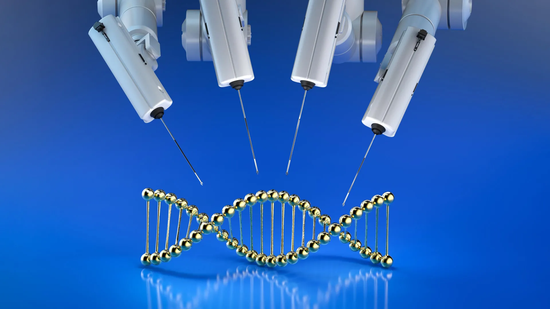
Some inherited diseases, including cystic fibrosis, hemophilia, and Tay Sachs disease, involve multiple genetic mutations within a person’s DNA. Even two individuals with the same condition may have different sets of mutations. Because of this…

For centuries, the moon has captivated our imagination, serving as a celestial companion and a canvas for scientific discovery. But beyond its silvery glow lies a rugged, crater-pocked world filled with fascinating features that tell the story…

Astronomers have discovered a peculiar ‘moon’ shadowing Earth as it moves through space. Known as a quasi-moon, it doesn’t orbit our planet directly but stays nearby as both travel around the Sun.
According to new research published in…